2018 WITec Paper Award for Outstanding Scientific Publications
Every year WITec, the Raman imaging company, recognizes three peer-reviewed publications that stand out in terms of originality and significance and feature data acquired with a WITec microscope. This year, scientists from universities and institutes in Bochum, Bayreuth and Minneapolis received the awards. Some 80 publications were submitted, all documenting the versatility of confocal Raman imaging for the investigation of materials.
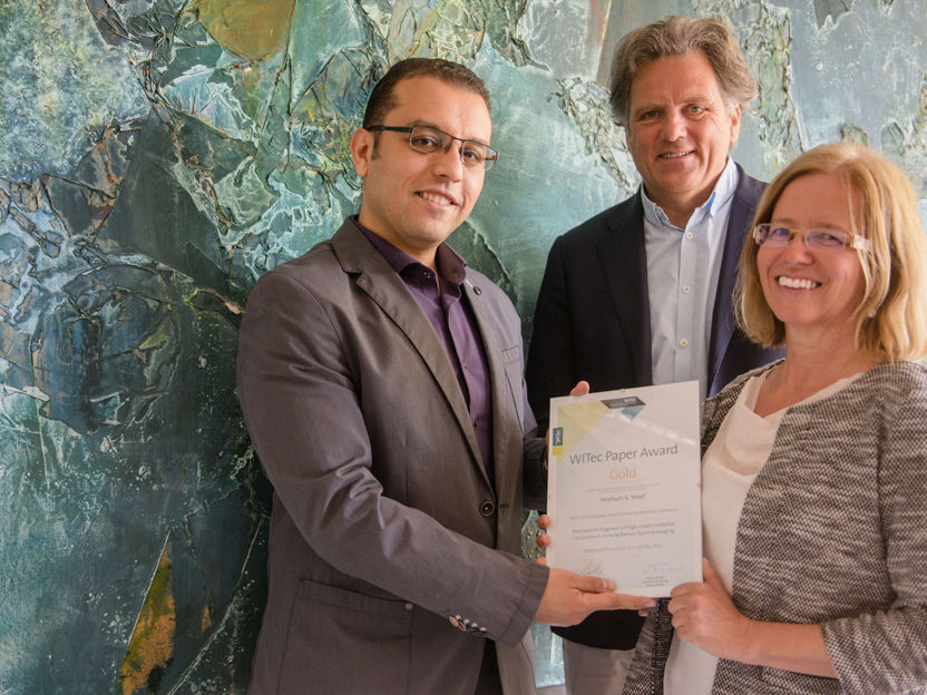
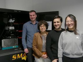
First author Hesham Yosef (left) and senior author Klaus Gerwert receive the winner’s certificate from WITec’s marketing assistant Karin Hollricher.
Copyright: RUB, Marquard
The Gold Paper Award
The Gold Paper Award goes to Hesham K. Yosef. With his colleagues from the Department of Biophysics, headed by Klaus Gerwert, and coworkers from other institutes at Ruhr-University Bochum (Germany) he developed a new, non-invasive approach based on Raman imaging for the detection of urothelial carcinoma cells in urine. Urothelial carcinomas constitute the majority of bladder tumors. Noninvasive, painless methods are urgently wanted for the diagnosis of this cancer and the monitoring of the treatment outcome. The scientists first identified differences in the Raman spectra of healthy and high-grade cancerous cells in urine. The most prominent differences in Raman bands indicated a decrease in the level of glycogen and an increase in the levels of fatty acids in the tumor cells, illustrating distinct metabolic changes. The scientists then developed and trained a classifier that could differentiate healthy from cancerous urothelial cells by their Raman spectra. A Raman band at 482 relative wavenumbers, indicating a massive loss of glycogen in tumor cells, emerged as a highly reliable marker. A first validation test on the urine of ten diagnosed and ten healthy patients was performed and the classifier diagnosed all samples automatically with 100% accuracy. First author of the publication, Hesham K. Yosef, says that he is convinced that, “The results will pave the way for spectral, noninvasive cytology of urine using Raman microspectroscopy.”
The Silver Paper Award
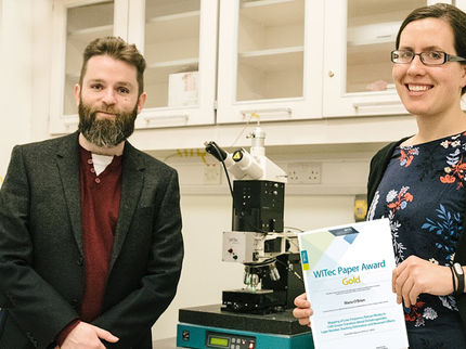
Mimicking biological structures with interesting functional features is a thriving field of research. Marvin Gernhardt, Holger Schmalz, Seema Agarwal and coworkers at the Chair of Macromolecular Chemistry II, University of Bayreuth, and the Bavarian Polymer Institute (Germany) were inspired by biological fibers and developed new fibers with distinct structures. They produced the fibers by side-by-side electrospinning of a thermo-responsive polymer and methacrylate-based copolymer. By changing the spinning solvents they fabricated side-by-side and coaxial bead-on-string fibers and analyzed these in detail with correlative Raman imaging, atomic force microscopy and scanning electron microscopy. The scientists concluded: “The work opens the way for the construction of heterostructured fiber morphologies based on different polymer combinations, offering high potential for applications as actuators, smart textiles, water management and catalysis.”
The Bronze Paper Award
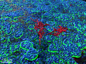
Guanglin Yu receives the Bronze Paper Award. He and his colleagues from the Departments of Mechanical Engineering and Biomedical Engineering at the University of Minnesota in Minneapolis (USA) analyzed, when and how ice crystals develop in cells during freezing. Cryopreservation is a common method used to store cells, however not all cells survive the procedure. It has been hypothesized that a high cooling rate leads to the death of cells because of intercellular ice formation. How that happens is still a matter of debate. The researchers used confocal Raman imaging to monitor the freezing response of lymphoblasts under various conditions during the cryopreservation process. Ice crystals, cell mass and protective substances could be easily imaged and identified by their Raman spectra. As a parameter of cell death, they used a Raman signal indicating the release of cytochrome c from mitochondria. The imaging study showed that only the formation of large intracellular ice crystals (> 2 μm) is deadly for the cells and that a mixture of glycerol and trehalose prevented cell death most efficiently at a cooling rate of 10°C per minute. “Raman spectromicroscopy provides a powerful tool for observing IIF [intracellular ice formation] and understanding its role in cell death during freezing, and enables the development, to our knowledge, of new and improved cell preservation protocols,” concluded the authors in their paper.
Original publication
GOLD: Hesham K. Yosef, Sascha D. Krauß, Tatjana Lechtonen, Hendrik Jütte, Andrea Tannapfel, Heiko U. Käfferlein Thomas Brüning, Florian Roghmann, Joachim Noldus, Axel Mosig, Samir F. El-Mashtoly and Klaus Gerwert; "Noninvasive diagnosis of high-grade urothelial carcinoma in urine by Raman spectral imaging"; Analytical Chemistry; 89, 6893 (2017).
SILVER: Marvin Gernhardt, Ling Peng, Matthias Burgard, Shaohua Jiang, Beate Förster, Holger Schmalz and Seema Agarwal; "Tailoring the morphology of responsive bioinspired bicomponent fibers"; Macromolecular Materials and Engineering; 303, 1700248 (2017).
BRONZE: Guanglin Yu, Yan Rou Yap, Kathryn Pollock and Allison Hubel; "Characterization intracellular ice formation of lymphoblasts using low-temperature Raman spectroscopy"; Biophysical Journal; 112, 2653 (2017).
Other news from the department science


Get the chemical industry in your inbox
From now on, don't miss a thing: Our newsletter for the chemical industry, analytics, lab technology and process engineering brings you up to date every Tuesday and Thursday. The latest industry news, product highlights and innovations - compact and easy to understand in your inbox. Researched by us so you don't have to.
Most read news


More news from our other portals


See the theme worlds for related content
Topic World Spectroscopy
Investigation with spectroscopy gives us unique insights into the composition and structure of materials. From UV-Vis spectroscopy to infrared and Raman spectroscopy to fluorescence and atomic absorption spectroscopy, spectroscopy offers us a wide range of analytical techniques to precisely characterize substances. Immerse yourself in the fascinating world of spectroscopy!