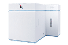
witec360 Raman Imaging Mikroskop
3D Raman microscopes with unequalled speed, sensitivity and resolution
Maximized signal intensity for the broadband range from 350 - 1100 nm excitation
Maximise efficiency and simplify your workflows with advanced automation options
Intelligent automation features lead to a higher productivity and sample turnover

Visualize and characterize every chemical detail
Take your research in any direction with the new benchmark for Raman imaging
The Oxford Instruments witec360 is a unique confocal Raman microscope capable of routinely performing 3D chemical Raman imaging while maintaining the highest measurement speed and spectral quality. This is particularly beneficial for applications in which the exact spatial representation of the chemical components on the surface or within the sample is important. Depth profiles, 3D image stacks or topographic Raman images can be easily created with maximum informational content. The local resolution is always dependent on the physical diffraction limit and has an approximate minimum of 200 nm. At the same time, high-speed measurements are possible in which up to 1300 spectra per second can be recorded.
With the most recent release of the new witec360 Raman microscope Oxford Instruments takes imaging automation to the next level. It offers eease-of-use and ultimate capability by automating hardware control and offering pre-configured measurement routines. This streamlines the experimental workflow and yields reproducible results with unrivaled speed, sensitivity and resolution.
A critical advantage of Oxford Instruments Raman technology is the flexible, high-throughput Hexalight Spectrometer. It is optimized for the a full broadband excitation wavelength range. As a result the user achieves a much higher throughput than with conventional spectrometers and can operate, for example, with a lower laser power, which is of particular benefit when investigating sensitive samples.
If the laser power is to be accurately measured and adjusted, the customer can be provided with the TruePower option, which can be used to determine the absolute laser power in increments of 0.1 mW. The values are recorded with the Raman data acquisition and can be retrieved later – important for internal documentation. The experiment can, of course, be repeated at a later time under precisely the same conditions.
Due to a modular design Raman microscopy can be correlated with AFM, SNOM or optical profilometry. With RISE microscopy, even correlative Raman-SEM images are possible.
The witec360 Raman imaging system offers several advantages for versatile research and development applications, without compromise in confocality while maintaining the highest spectral performance and speed and providing detailed 3D analyses of different samples. Straightforward operation is ensured by new innovative operating concepts. Raman imaging is therefore a flexible and versatile tool for chemical imaging and analysis.
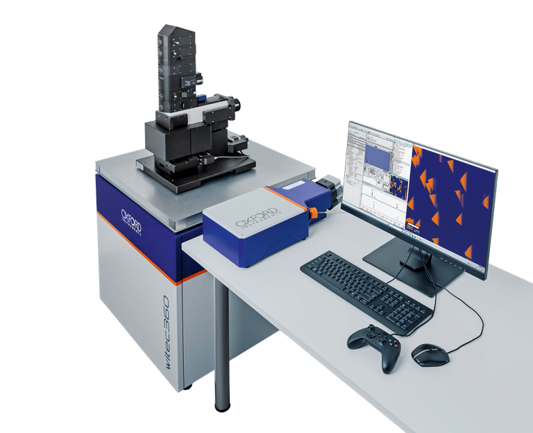
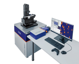
1
witec360 Raman Imaging System
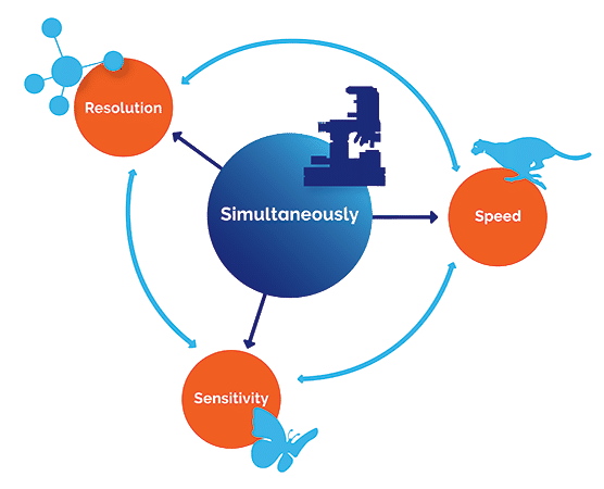
2
Speed-Sensitivity-Resolution: Simultaneously
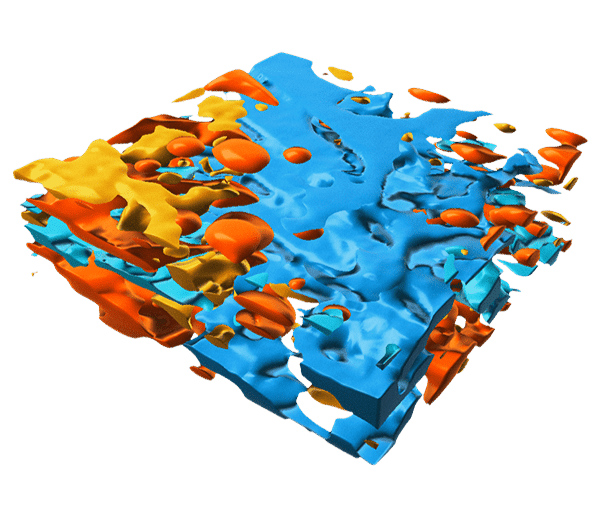
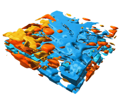
3
3D Raman image of a cosmetic emulsion. (30 x 30 x 10 µm³).
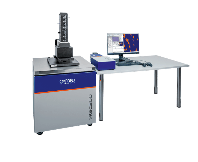
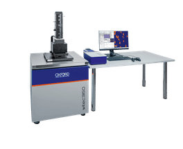
4
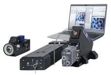
witec360 with the new Hexalight Broadband Spectrometer
Request information about witec360 Raman Imaging Mikroskop now
Raman microscopes: witec360 Raman Imaging Mikroskop
3D Raman microscopes with unequalled speed, sensitivity and resolution
Product classification witec360 Raman Imaging Mikroskop
Product categories
Applications
Manufacturers of similar products


Find more Raman microscopes and related products
Find witec360 Raman Imaging Mikroskop and related products in the theme worlds
Topic World Spectroscopy
Investigation with spectroscopy gives us unique insights into the composition and structure of materials. From UV-Vis spectroscopy to infrared and Raman spectroscopy to fluorescence and atomic absorption spectroscopy, spectroscopy offers us a wide range of analytical techniques to precisely characterize substances. Immerse yourself in the fascinating world of spectroscopy!