First Science from the Compact Light Source: A miniature synchrotron for your home lab
In 2004 Lyncean Technologies announced the construction of the Compact Light Source (CLS), a miniature synchrotron which uses inverse Compton scattering to produce high-intensity, tunable, near-monochromatic x-ray beams. The CLS was designed to bring state-of-the-art protein structure determination to the home laboratory — but it has also promised to have a broad impact across the spectrum of x-ray science.
Today, at the 39th Winter Colloquium on the Physics of Quantum Electronics, Ronald Ruth, Ph.D., president of Lyncean Technologies, announced that the CLS has started delivering on this promise by achieving three key milestones using its unique x-ray beam: First scientific publication from the CLS featured by the Journal of Synchrotron Radiation on its January 2009 cover; first micro-tomographic images from the CLS; and first protein crystallography data set from the CLS.
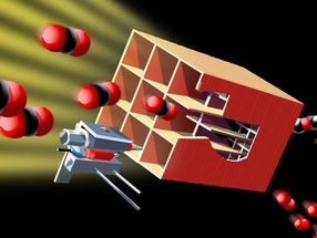
The Compact Light Source prototype effort was funded by the National Institute of General Medical Sciences (NIGMS) Small Business Innovation Research (SBIR) program as an advanced technology related to the NIGMS Protein Structure Initiative (PSI). The CLS technology is based on an electron beam stored in a miniature storage ring colliding repeatedly with an opposing infrared light pulse stored in a high-finesse cavity. Each collision produces x-rays through inverse Compton scattering. The entire x-ray source fits in a 10x25 ft room similar in size to those used for Magnetic Resonance Imaging in medical clinics.
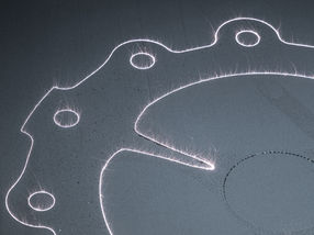
The first scientific publication using the CLS x-ray beam employs a technique called Differential Phase Contrast Imaging (DPCI) developed by Professor Franz Pfeiffer and collaborators at the Paul Scherrer Institute in Switzerland. DPCI uses a pair of micron-scale gratings to create two images, one sensitive to the phase of the x-ray wave front and the other sensitive to the local scattering power. This technique has been primarily developed at x-ray beam lines in large synchrotrons, and it relies on a small point-like x-ray source to achieve the coherence necessary for the fringes. The Compact Light Sources has an x-ray beam that is a perfect match for the technique.
In the second round of the Protein Structure Initiative, further CLS development was included in the Accelerated Technology Center for Gene to 3D Structure (ATCG3D) supported by both NIGMS and the National Center for Research Resources (NCRR). "The focus of the CLS development has been towards protein crystallography," said Professor Ruth, "and with the ATCG3D Beta CLS we had the opportunity to improve performance and reliability. We now have the Beta CLS nearly ready for installation and further development."
With the publication of the first scientific results and the collection of the first set of crystallography data using the CLS, the instrument has demonstrated its potential to have a broad impact on biomedical research," said Jeremy M. Berg, Ph.D., director of NIGMS. "The wide availability of an intense, tunable x-ray tool could transform numerous fields of research by improving access to a key resource."
Other news from the department research and development


Get the chemical industry in your inbox
From now on, don't miss a thing: Our newsletter for the chemical industry, analytics, lab technology and process engineering brings you up to date every Tuesday and Thursday. The latest industry news, product highlights and innovations - compact and easy to understand in your inbox. Researched by us so you don't have to.