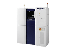
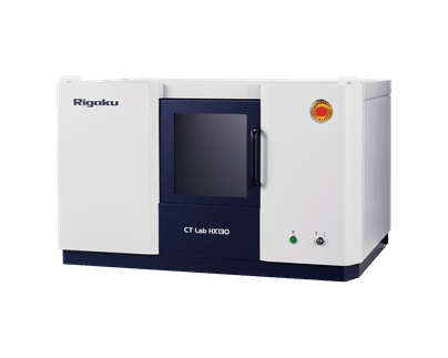
CT Lab HX
Rigaku CT Lab HX – Benchtop Micro Computed Tomography System
Key Features
- Powerful 130 kV X-ray source
- 2.1 µm voxel resolution
- Wide 200 mm field of view (FoV)
- Temporal resolution – down to 18 sec
- Max. sample size - 200 mm Φ x 270 mm high
- Flexible design to cover high-resolution to large FOV
- Compact benchtop design
- Auto sample changer available
Powered by a class-leading 130 kV X-ray source, the Rigaku CT Lab HX is a compact and versatile micro-CT (computed tomography) system suited to non-destructive 3D imaging applications spanning materials science, life science, and geology in both research and industry. Its flexible geometry realizes 2.2 µm voxel resolution and 200 mm field of view (FOV) on high-resolution and large FOV sides, respectively.
While other imaging techniques might see only the surface or require sectioning of the sample, using the Rigaku CT Lab HX, you can easily look inside your sample non-destructively. Study fiber orientation of composites, internal organs, and skeletal structure of insects and small animals, or check for dimensional accuracy or detect flaws, pores, cracks, or other undesirable features to best ensure the integrity of your masterpiece.
A key feature of the CT Lab HX is the ability to vary the SOD (Source-to-object distance) and the SDD (source-to-detector distance), allowing you to optimize your geometry for high resolution to a wide FOV. Fast scan times allow high throughputs as well as the ability to perform time-resolved studies.
Applications
The Rigaku CT Lab HX is ideally suited to applications in:
- Pharmaceuticals – Reveal defects in pills, tablets, and capsules such as cracks, voids, coating non-uniformity or delamination, aggregates, phase change, and degradation, which can affect the efficacy and performance of your drugs.
- Foams and composites – CT provides the unique ability to image complex structured materials in 3D, allowing the determination of structure, fiber, void, cell, or pore distribution and the ability to quantify over large volumes. This is not possible using other microscopy techniques, especially non-destructively.
- Metrology – With the rapid growth of 3D printing and additive manufacturing, CT is following suit as a valuable technique to check the dimensional accuracy of finished products and compare them to design or CAD drawings. While more traditional techniques like CMM (Co-ordinate Measuring Machines) only measure a limited number of points on a surface, CT can image the entire volume, including the internal structures, voids, pores, cracks, inclusions, and other undesirable features, providing insights to the design optimization and failure analyses. Conversely, CT can also help reverse manufacture existing components.
- Life science – CT allows imaging plants, animals, and insects with next to no sample preparation. It also allows imaging of internal organs and structures without sectioning the subject. Time-resolved studies can reveal processes such as germination, metamorphosis of insects, or infestation of crops, which are otherwise hidden from sight.
- Geology – CT is used extensively in exploration geology to determine properties such as phase fraction of rocks, porosity or pore network in sandstones, or oil and water distribution in drill cores. CT images can be used for Digital Rock Physics to help better focus expensive drilling programs.
- Paleontology – CT can examine fossils and other historically significant artifacts non-destructively to see what lies below the surface, potentially revealing hidden features, while the original specimen remains intact for future use.
- Food – CT is applied to a wide range of food types, including fruits, vegetables, grains, meats, and processed foods. CT can be used to determine fat, protein, and water distribution, to examine food coatings, salt distribution, and texture of dairy products. Time-resolved studies can also be used to evaluate baking/cooking processes.