TU Wien develops a microscope for ultra-sensitive samples
How can you produce the best possible image in a microscope without destroying the sample? A new trick enables gentle imaging with maximum image quality.
Advertisement


Everyone who ever took a photo knows the problem: if you want a detailed image, you need a lot of light. In microscopy, however, too much light is often harmful to the sample – for example, when imaging sensitive biological structures or investigating quantum particles. The aim is therefore to gather as much information as possible about the object under observation with a given amount of light.
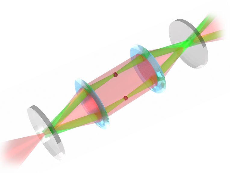
The new type of microscope makes light travel in a circle. That way it can interact multiple times with the sample.
© TU Wien
In a collaboration with the University of Vienna and the University of Siegen (Germany), researchers at TU Wien have now developed a novel trick to achieve this: they store the light in a resonator in which the sample is also located. This allows them to obtain a clearer signal than with other methods. The technique has now been presented in the journal Nature Scientific Reports.
Better signal through multiple light scattering
“In a normal microscope, the light hits the sample once and then enters a lens,” says Maximilian Prüfer, who led the study as part of his Esprit fellowship from the FWF at the Atomic Research Institute of TU Wien. “In our microscope, we place the sample in an optical resonator – between two mirrors.”

To turn this resonator into a microscope, the team developed an unusual experimental setup with additional lenses: after the light beam has passed through the sample, it is guided in a circle and hits the sample again. “Now the sample is illuminated again, but not with a normal, uniform beam of light as in the beginning, but with a beam of light that already contains the image of the sample, so to speak,” explains Oliver Lueghamer (TU Wien), who built the microscope as part of his master's thesis.
Similar to a stamp that is pressed several times on the same spot, producing a clearly visible image even with faint ink, the image of the sample becomes clearer and clearer as it completes several rounds in the microscope.
Both theoretical calculations, which were developed in collaboration with Thomas Juffmann (University of Vienna) and Stefan Nimmrichter (University of Siegen), and experiments show that this method provides more information than other microscopy techniques at a given light intensity. “The key figure is the signal-to-noise ratio,” explains Maximilian Prüfer. “This ratio is better here than with other methods due to multiple scattering with the same disturbance of the sample.”
Stable even with minor disturbances
However, the practical suitability of the new method also depends on how susceptible it is to disturbances: “When using optical resonators, as we do, it is often important to keep their length extremely constant,” says Maximilian Prüfer.
“Normally, you have to go make a great effort to ensure that the distance between the two mirrors varies only minimally, otherwise the desired effect is lost. With our method, however, this is not the case.”
The distance between the mirrors can also show a certain instability without the enhancement disappearing. “This is important because it means that the method not only works in theory, but can also be used in practice with manageable effort,” says Prüfer.
One of the goals of the new microscopy technique is to image ultra-cold Bose-Einstein condensates and thereby study their quantum physical behaviour.
Original publication

Other news from the department science


These products might interest you

alpha300 R by WITec
3D Raman microscopes with unequalled speed, sensitivity and resolution
Visualize and characterize every chemical detail
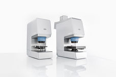
LUMOS II by Bruker
FT-IR microscopy in the fast lane - the LUMOS II
One infrared microscope for all
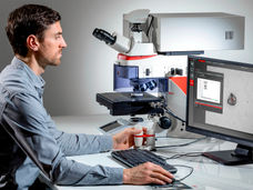
DM8000 M & DM12000 M by Leica
See More, Detect Faster
High-throughput Inspection Systems
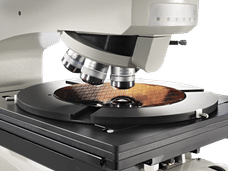
HYPERION II by Bruker
FT-IR and IR laser imaging (QCL) microscope for research and development
Analyze macroscopic samples with microscopic resolution (5 µm) in seconds