To use all functions of this page, please activate cookies in your browser.
My watch list
my.chemeurope.com
my.chemeurope.com
With an accout for my.chemeurope.com you can always see everything at a glance – and you can configure your own website and individual newsletter.
- My watch list
- My saved searches
- My saved topics
- My newsletter
Near-field scanning optical microscopeNear Field Scanning Optical Microscopy (NSOM/SNOM) is a microscopic technique for nanostructure investigation that breaks the far field resolution limit by exploiting the evanescent near field diffraction. This is done by placing the detector very close (<< λ) to the specimen surface. This allows for the surface inspection with high spatial, spectral and temporal resolving power. As in optical microscopy, the contrast mechanism can be easily adapted to study different properties, such as refractive index, chemical structure and local stress. Dynamic properties can also be studied at a sub-wavelength scale using this technique. Product highlight
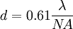
History of NSOME.H. Synge, an Irish scientist, is given credit for conceiving and developing the idea for an imaging instrument that would image by exciting and collecting diffraction in the near field. His original idea, proposed in 1928, was based upon the usage of intense nearly planar light from an arc under pressure behind a thin, opaque metal film with a small orifice of about 100nm. The orifice was to remain within 100nm of the surface, and information was to be collected by point-by-point scanning. He foresaw the illumination and the detector movement being the biggest technical difficulties. [1] [2][3]. J.A. O’Keefe also developed similar theories in 1956 without knowledge of Synge’s earlier works. He thought the moving the pinhole or the detector when it is so close to the sample would be the most likely issue that could prevent the realization of such an instrument.[4] It was Ash and Nichols who, in 1972, first broke the Abbe’s diffraction limit using 3cm radiation. A line grating was resolved with a resolution of λo/60 in this work. [5] It was twelve more years before the first papers that used visible radiation for near field scanning were published by Pohl et al., and Lewis et al. Both these works involved the use of a subwavelength metal coated optical aperture at the tip of a sharp pointed probe, and a feedback mechanism to maintain a constant distance of a few nanometers between the sample and the probe [6][7]. Resolutions as low as 25nm (about λo/20) were achieved in the work by Pohl et al[citation needed]. TheoryAccording to Abbe’s Theory of Image Formation, developed in 1873, the resolving capability of an optical component is ultimately limited by the spreading out of each image point due to diffraction. Unless the aperture of the optical component is large enough to collect all the diffracted light, the finer aspects of the image will not correspond exactly to the object. The minimum resolution (d) for the optical component are thus limited by its aperture size, and expressed by the following relationship: Here, λo is the vacuum wavelength; NA is the numerical aperture for the optical component (usually 1.3-1.4 for modern objectives). Thus, the resolution limit is usually around λo/2 for conventional optical microscopy. [8] This treatment only assumes the light diffracted into the far-field that propagates without any restrictions. NSOM makes use of evanescent or non propagating fields that exist only near the surface of the object. These fields carry the high frequency spatial information about the object and have intensities that drop off exponentially with distance from the object. Because of this, the detector must be placed very close to the sample in the near field zone, typically a few nanometers. As a result, near field microscopy remains primarily a surface inspection technique. The detector is then rastered across the sample using a piezoelectric stage. The scanning can either be done at a constant height or with regulated height by using a feedback mechanism. [9]
Modes of OperationAperture and Apertureless OperationNSOM can be operated in both, an aperture and a non-aperture, mode. As illustrated, the tips used in the apertureless mode are very sharp and do not have a metal coating. Though there are many issues associated the apertured tips (heating, artifacts, contrast, sensitivity, topology and interference amongst others), aperture mode remains more popular. This is primarily because apertureless mode is even more complex to set up and operate, and is not understood as well. There are five primary modes of apertured NSOM operation and four primary modes of apertureless NSOM operation. The major ones are illustrated in the next figure.
Feedback MechanismsFeedback mechanisms are usually used to achieve high resolution and artifact free images since the detector must be positioned within a few nanometers of the surfaces. Some of these mechanisms are:
ContrastIt is possible to take advantage of the various contrast techniques available to optical microscopy though NSOM but with much higher resolution. By using the change in the polarization of light or the intensity of the light as a function of the incident wavelength, it is possible to make use of contrast enhancing techniques such as staining, fluorescence, phase contrast and differential interference contrast. It is also possible to provide contrast using the change in refractive index, reflectivity, local stress and magnetic properties amongst others. [9][10] Instrumentation and Standard SetupThe primary components of an NSOM setup are the light source, feedback mechanism, the scanning tip, the detector and the piezoelectric sample stage. The light source is usually a laser focused into an optical fiber through a polarizer, a beam splitter and a coupler. The polarizer and the beam splitter would serve to remove stray light from the returning reflected light. The scanning tip, depending upon the operation mode, is usually a pulled or etched optical fiber coated with metal except at the tip or just a standard AFM cantilever with a hole in the center of the pyramidal tip. Standard optical detectors, such as Avalanche Photo Diode (APD), Photomultiplier Tube (PMT) or CCD, can be used. Highly specialized NSOM techniques, Raman NSOM for example, have much more stringent detector requirements. A standard setup for an apertureless reflection-back-to-the-fiber NSOM system is illustrated in the next figure. [10] Near Field SpectroscopyAs the name implies, information is collected by spectroscopic means instead of imaging in the near field regime. Through Near Field Spectroscopy (NFS), one can probe spectroscopicaly with subwavelength resolution. Raman SNOM and fluorescence SNOM are two of the most popular NFS techniques as they allow for the identification of nanosized features with chemical contrast. Some of the common near field spectroscopic techniques are:
ArtifactsNSOM is particularly vulnerable to artifacts that are not from the intended contrast mode. The most common root for artifacts in NSOM are:
Limitations of NSOM
References
See also
|
|||||||||||||
| This article is licensed under the GNU Free Documentation License. It uses material from the Wikipedia article "Near-field_scanning_optical_microscope". A list of authors is available in Wikipedia. | |||||||||||||