New technique paves way for imaging individual molecules
Pioneering experiment exploits quantum properties of X-ray light
Advertisement


An international research team has succeeded for the first time in using X-rays for an imaging technique that exploits a particular quantum property of light. As the researchers describe in their work just published in the journal Physical Review Letters, this technique could enable imaging of non-crystallized macromolecules.
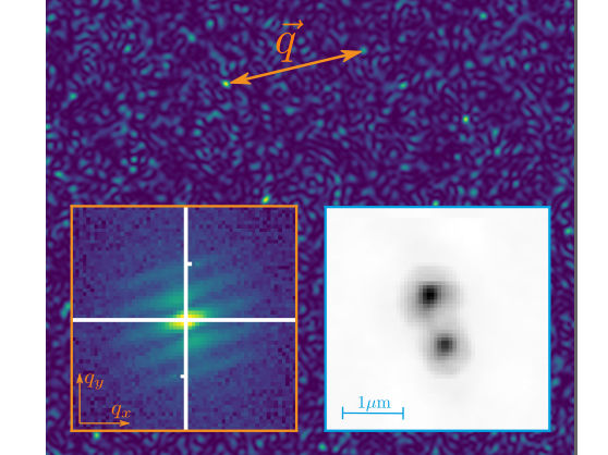
Light emitted from independent luminous sources, such as fluorescence from atoms, creates waves at random times and thus with random phases. If measured within its coherence time, this light will interfere and produce a speckle pattern such as shown in the background image. This pattern is not stationary and the sum of many such patterns will average to a uniform distribution. However, if instead pair correlations are calculated from each pattern and then summed, the random phases will average away to leave a map of the spatial frequency content (q vector) of the sources. The sum of over 58 million correlations of X-ray fluorescence snapshots is shown in the left insert, which was analysed by methods of coherent diffractive imaging to produce a high-resolution image of the source—here two illuminated spots in a spinning copper disk.
DESY, Fabian Trost
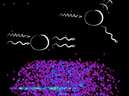
The research team, led by Henry Chapman, leading scientist at DESY and professor at Universität Hamburg, used very intense X-ray pulses from the X-ray free-electron laser European XFEL to generate fluorescence photons that arrived almost simultaneously at the detector – within a time window shorter than a femtosecond (a quadrillionth of a second). By computing photon-photon correlations of the X-ray fluorescence emitted by the illuminated copper atoms, images of the emission could be obtained.
The structures of materials and macromolecules are usually determined at the atomic scale using X-ray crystallography. While that technique relies on coherent X-ray scattering, incoherent processes like fluorescence emission can dominate even though they do not usefully contribute to the diffraction measurement. Instead, they add a featureless fog or background to the measured data.
But already in the 1950s, two British astronomers demonstrated that it is indeed possible to extract structural information from such light emitted by self-luminous sources – in their case from stars. The method of Robert Hanbury Brown and Richard Twiss – called intensity interferometry – opened a new door into the understanding of light and started the field of quantum optics.
Recently, scientists from the University of Erlangen, the Max Planck Institute for the Structure and Dynamics of Matter and DESY proposed that intensity interferometry could be adapted for atomic-resolution imaging using X-ray fluorescence. The challenge in extending this idea to X-rays is that the coherence time of the photons, which dictates the time interval available to perform photon-photon correlations, is extremely brief. It is set by the radiative decay time of the excited atom, which for copper atoms is about 0.6 femtoseconds.
Now the group, together with scientists from Uppsala University and the European XFEL have overcome that challenge by using femtosecond-duration XFEL pulses from that facility to initiate X-ray fluorescence photons within the coherence time. They generated a source consisting of two fluorescing spots in a foil of copper and measured the fluorescence on a million-pixel detector placed eight metres away. “For this pioneering experiment we collected over three petabytes of data, the largest ever for an experiment at the European XFEL,” explains main author Fabian Trost from the Center for Free-Electron Laser Science (CFEL) at DESY. “However, since the signal scales with the square of intensity, we see that it should be possible to reduce this substantially in our future experiments.”
Only about 5000 photons were detected on each illumination pulse, and the cumulative sum over 58 million shots gave just a featureless uniform distribution. However, when they instead summed photon-photon correlations, a fringe pattern emerged, similar to the famous double-slit experiment. This fringe pattern is the smoking gun that indicates interference of separate X-ray photons. The fringe pattern was then analyzed as if from a coherent wave field to reconstruct an image of the fluorescent source, consisting of two well-separated spots.
“Although the idea of interference of independent waves within the coherence time can be understood classically and can be noticed in the interference of radio stations for example, with X-rays we are very much dealing with high energy quanta”, says Chapman, who is a researcher at the Cluster of Excellence CUI: Advanced Imaging of Matter. “Each fluorescence photon is born within a single atom, and these photons are then located at specific pixels of our detector. However, these photons carry hidden information that is only revealed when their higher-order photon-photon correlations are examined.”
The scientists now hope to combine this novel method with diffraction to image single molecules. The fluorescence will provide sub-structures specific to particular atoms and even particular chemical states of those atoms, which may help unravel the functioning of important enzymes such as involved in photosynthesis.
Original publication


Other news from the department science


