From Microscopy to Nanoscopy
Photoswitchable rhodamine amides for high-resolution optical 3D far-field microscopy
Advertisement


Layer-by-layer light microscopic nanoscale images of cells and without having to prepare thin sections? A team led by Stefan Hell and Mariano Bossi at the Max Planck Intstitute for Biophysical chemistry in Göttingen is now leading the way with a technique called optical 3D far-field microscopy - with nanoscale resolution, good signal-to-noise ratio, and relatively short exposure times. The secret of their success lies in special photoswitchable fluorescence dyes, the researchers report to the journal Angewandte Chemie. These rhodamine amides make it possible to obtain highly resolved 3D images of transparent fluorescence-marked samples such as living cells.
Until fairly recently, the resolution of light microscopes was limited by the wavelength of the light.There are non-optical methods, such as electron microscopy, but light microscopy is still the only way to observe the interior of whole, or even living, cells. The use of fluorescent dyes makes it possible to selectively obtain images of individual cell components, for example, proteins. Today, the wavelength dogma is overcome. Hell received the German Future Prize in 2006 for the first concept breaking the wavelength barrier the stimulated emission depletion (STED) microscope . Molecules are transferred from a "dark" (non-fluorescent) to a "bright" (fluorescent) excited energy state-with a spacial sharpness far beyond those 200 nanometers.
Now the german team is demonstrating the power of another concept. They use molecules that are not only transferred but can be "switched" from "fluorescent" to "non-fluorescent" and back. In contrast to the STED and other related methods of the team, only separate, isolated marker molecules are randomly switched on at the same time. Their fluorescence is registered, and then they get switched off again automatically. In this way, the simultaneously fluorescing (switched on) markers are farther apart from each other than the minimum distance that the microscope can resolve. This is only possible using switchable molecules that emit many photons, one after the other, when switched on. If these photons are captured with a camera, the centers of the individual fluorescing dots can be distinguished. After the exposure, the molecule becomes dark again (switches off), allowing further, neighboring molecules to be photographed. This process is repeated many times, until many dots become a picture. The full distribution can be reconstructed - at a resolution not limited by the wavelength of light.
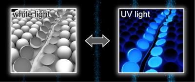
The researchers have now found a class of substances that fulfill all the requirements of this technique: rhodamine amides. At the core of these molecules lies a system of five rings. In this form, the compound is colorless and does not fluoresce. Irradiation with light induces an isomerization in which one of the rings is opened. This form of the molecule is red and can be excited several times.
Most importantly: rhodamine amides can be switched on by either a UV photon or two photons in the red part of the spectrum. This two-photon excitation can be focused onto a thin plane, which allows biological samples to be photographed layer by layer. The individual images can then be reconstructed into a single multilayer image. The resolution reached in the focal plane is far beyond the diffraction barrier (10-30 nm).
Original publication: Stefan W. Hell et al.; "Photochromic Rhodamines Provide Nanoscopy with Optical Sectioning"; Angewandte Chemie International Edition 2007, 46, No. 33, 6266-6270.


Other news from the department science


