New Microscopy tool for energy research
Technique allows scientists to explore the properties of photoelectrodes more precisely
Advertisement


Researchers at Helmholtz-Zentrum Hereon, the Helmut Schmidt University, the Lawrence Berkeley National Lab, and the Helmholtz-Zentrum Berlin have developed a promising approach for detecting voltage changes on the surface of photoelectrodes utilizing a newly developed automated data analysis method. The method presented in the journal PRX Energy enables so-called “Kelvin probe force microscopy (KPFM)” measurements in the millisecond range. It works by extracting information contained in each pixel of a KPFM image, which was previously not possible. The knowledge gained in this way can contribute to the development of more efficient and stable materials for photoelectrochemical cells (PECs).
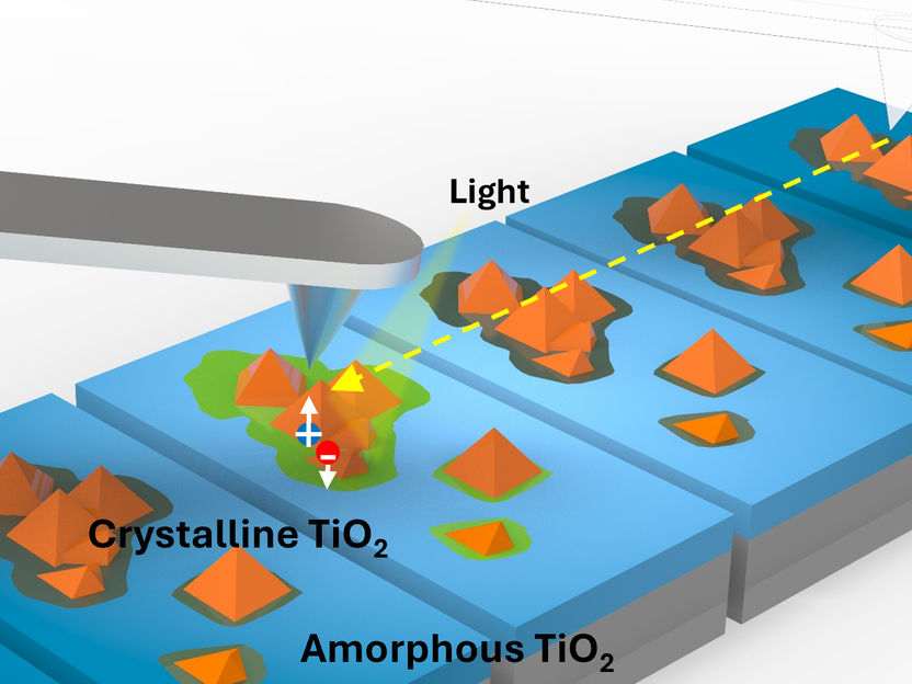
Schematic representation of the technique presented in the paper. The authors developed an analysis tool to obtain MS resolution using KPFM.
Hereon/Dr. Sehun Seo
PECs are cells that convert light into chemical energy and support the sustainable production of hydrogen and other chemicals such as fuels. Photoelectrodes, the central components of PECs, are light-sensitive and consist of semiconductors. In PECs, the semiconductors absorb light and thereby generate charge carriers that ultimately drive the chemical reactions. Despite their promising potential, these systems have not yet become established. The efficiency with which they can convert sunlight into hydrogen is still lower than theoretically predicted. In addition, their performance deteriorates significantly over time.
To enhance efficiency and stability over time, researchers need reliable instruments. Such as high-resolution microscopy to study the underlying structure and its light-related (optoelectronic) properties.
An advanced microscopy instrument for energy research
The team of researchers has devised the technology that can help here. It allows to study the interplay between the local morphology of a photoelectrode (i.e. the structure of small areas on its surface) and its charge transport dynamics (i.e. how well electrons and holes move in a material).
The new approach, presented by Prof. Dr. Francesca M. Toma, the lead author of the paper and head of the Institute of Functional Materials for Sustainability, works by measuring tiny voltage changes that occur in small areas of a photoelectrode`s surface when it is exposed to light. The researchers have used their technique in collaboration with the Lawrence Berkeley National Lab to study titanium dioxide (TiO2), a semiconductor material commonly used to make photoelectrodes.
"With our new automated data analysis method, we can track tiny voltage changes on the surface of a photoelectrode down to the millisecond in a way that has never been possible before," explains Dr. Mauricio Schieda, lead author of the paper. "Titanium dioxide is a simple system that allowed us to develop this approach. And also to show that it is possible to track the movement of charges under light. This brings us one step closer to improving solar fuel technologies."
“I was excited to understand how the tiny morphology of a photoelectrode affects how charges move when exposed to light,” says Maryam Pourmahdavi, PhD student at Helmut Schmidt University, who is working on the project at the Hereon Institute and is first author of the paper. “This knowledge is the key to designing photoelectrochemical cells that are more efficient and long-lasting.”
Informing the design of future photoelectrodes
With the developed technique, the researchers gained new insights into the connection between the structure of small areas on a photoelectrode and its charge transport dynamics. The same approach could soon be used to study materials other than TiO2, potentially contributing to the development of more effective photoelectrodes for PECs.
“This work originates after years of progress by our group and the community in advancing atomic force microscopy techniques to investigate photoelectrochemical materials combined with more recent data science approaches that allow more and more information to be extracted out of a simple image,” says Prof. Dr. Toma. “Now that we have demonstrated the potential of the technique on a model system such as TiO2, we are ready to study many more materials and discover even more efficient ones.”
Original publication
Maryam Pourmahdavi, Mauricio Schieda, Ragle Raudsepp, Steffen Fengler, Jiri Kollmann, Yvonne Pieper, Thomas Dittrich, Thomas Klassen, Francesca M. Toma; "Correlating Local Morphology and Charge Dynamics via Kelvin Probe Force Microscopy to Explain Photoelectrode Performance"; PRX Energy, Volume 4, 2025-6-9


Other news from the department science


