A new way to record nanoparticles
Two-colour X-ray pulses capture the same sample at two moments in time
Advertisement


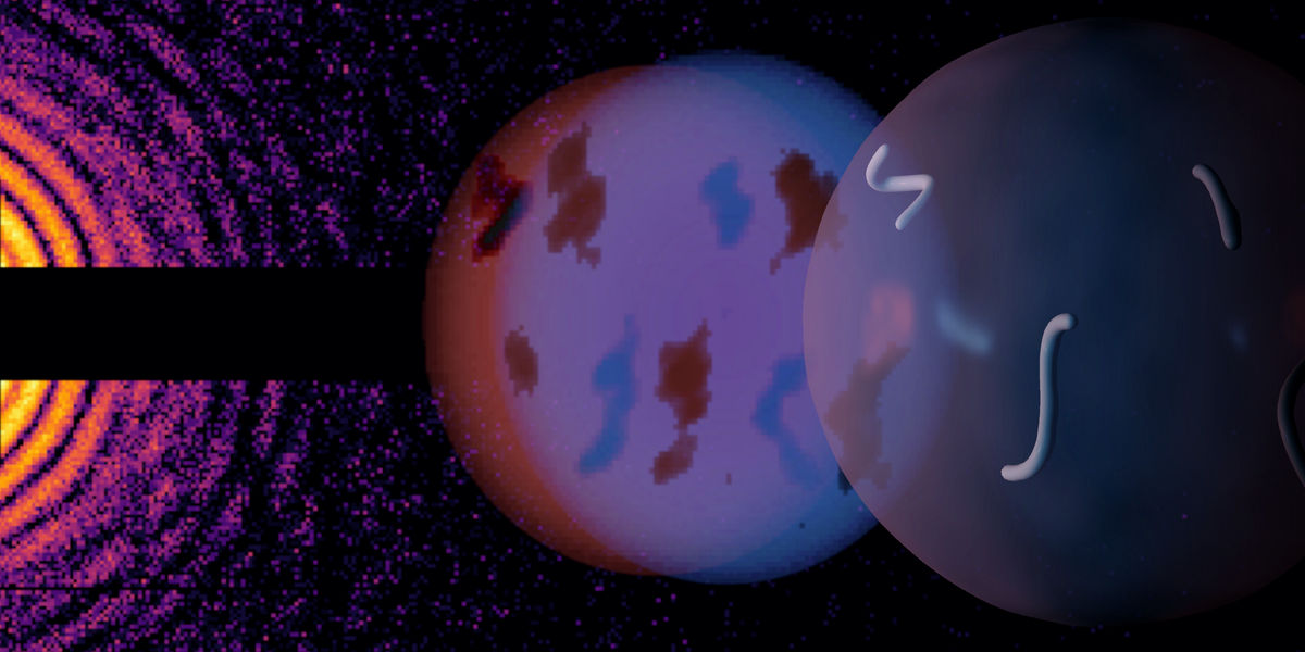
One big dream of ultrafast science has been to watch changes in nanoparticles or biomolecules on their natural timescale. Experimentally, this can be realized by taking two snapshots of the same object only femtoseconds (millionth of a billionth of a second) apart. However, no detector is fast enough to record the two snapshots separately—they end up on top of each other in a single image. Scientists now employed two complementary methods to disentangle two diffraction patterns captured by the same X-ray detector. The results by an international team of researchers have been published their results in two separate articles in Nature Communications.
A new capability of the European XFEL has recently enabled a big step towards the realization of nanoscale movies: the X-ray laser can produce subsequent flashes of two different colours, that are bright enough to image a particle twice within femtoseconds. Both methods to disentangle two images take advantage of these different colours or photon energies: one method exploits the capability of the detector to discern energy levels for each individual pixel, while the other uses mathematical reconstructions. Its inventors call the latter method Dichography. One of the leading scientists compares the technique to an extreme high-speed camera: "To my knowledge, these are the fastest nanoscale movies ever recorded, if by movie we mean multiple frames of the same object," says Alessandro Colombo from the Department of Physics at ETH Zurich in Switzerland.
“This opens the door to observe rapid changes in nanoparticles, comparable to studies that have been possible with small molecules for some time now,” says Yevheniy Ovcharenko, scientist at the Small Quantum Systems (SQS) instrument at European XFEL and principal investigator of the study. “These nanoparticles are less uniform than individual molecules, which makes it necessary to observe processes using the very same sample.” Experiments on single molecules that follow chemical processes usually rely on series of observations of different samples at varying time delays after a trigger event. That approach does not produce precise results for particles that can differ in size, shape, orientation, or internal structure. With two X-ray captures of the same particle, taken at a variable time delay, researchers can begin to follow a unique event directly, such as an expanding nanoplasma, a fragmenting cluster, or a structural rearrangement triggered by light. “We are at a starting point for a new type of study,” adds Marcel Mudrich of the University of Kassel, who proposed the experiment together with his SQS colleague. “We now have free- electron lasers that can produce pairs of X-ray pulses with different ‘colours’, we have detectors that distinguish between images based on the colour of the scattered X-ray light, and we have the analysis tools to reconstruct the shape of individual particles from the recorded X-ray snapshots.”
To develop and prove the feasibility of the colour-separation and Dichography methods, the scientists used the SQS instrument at European XFEL. Two sections of the facility’s undulators, X-ray light sources with a total length of 120 meters, were tuned to different X-ray photon energies (about 1.0 and 1.2 kiloelectronvolts), or “colours,” creating two X-ray pulses. The second X-ray pulse was emitted tens to hundreds of femtoseconds after the first. Both pulses were then focused onto free-flying helium nanodroplets and the scattered X-rays were recorded on a pnCCD detector. This device measures the charge created by incoming photons, which makes it possible to estimate their energy at each pixel. By analysing the charge deposited in each pixel and small clusters of pixels, the team could assign many detected photons to the first or second X-ray pulse. “This is more complicated than it sounds,” says Michael Meyer, lead scientist and group leader at the SQS instrument and one of the authors of the publications, “because we have a lot of photons. One pixel might have collected one photon from the first pulse and multiple photons from the second pulse. In addition to that, one photon can also illuminate multiple pixels.” Linos Hecht, Ph.D. candidate at ETH Zurich and first author of the publications, underlines that “the method is very flexible, because it doesn’t rely on any specific sample property. Furthermore, it works best in sparsely illuminated areas of the detector at large scattering angles, where the most precious high-resolution information is stored.”
That pixel-level colour separation provided the experimental proof of principle. The second step was more ambitious: instead of sorting detected photons by colour, the researchers aimed to recover two actual images of the sample from one mixed diffraction pattern. That is the idea behind Dichography, developed in a companion study. The method uses iterative phase- retrieval algorithms to search for two separate structures whose diffraction intensities add up to the measured signal. In the European XFEL data, the team reconstructed two time-separated views of a helium nanodroplet, which contained xenon particles, from single overlapping patterns at delays of 50 and 750 femtoseconds. The two-frame movies showed that the embedded xenon nanoclusters looked essentially unchanged: while intense light can cause particles to explode, the process had barely begun within the observed time span. The experiments and accompanying simulations also showed that the problem remains hard: Dichography works only when the data are bright enough and the two colour contributions are reasonably balanced. “I’m positive that continued development at facilities like European XFEL will make the limitations less and less of an issue and our method more and more applicable,” Colombo says. “We’re setting out on a new scientific adventure, making the dream of filming ultrafast structural changes in individual nanoparticles finally come true.”
Original publication
Linos Hecht, Andre Al Haddad, Björn Bastian, et al.; "Dichography: two-frame ultrafast imaging from a single diffraction pattern"; Nature Communications, Volume 17, 2026-6-24
Linos Hecht, Yevheniy Ovcharenko, Asbjørn Ø. Lægdsmand, et al.; "Model-free pattern separation of two-color ultrafast X-ray diffraction"; Nature Communications, 2026-9-3


Other news from the department science













Most read news









More news from our other portals






















See the theme worlds for related content
Topic World Particle Analysis
Particle analysis methods allow us to study tiny particles in various materials and reveal their properties. Whether in environmental monitoring, nanotechnology or the pharmaceutical industry, particle analysis gives us a glimpse into a hidden world where we can decipher the composition, size and shape of particles. Experience the fascinating world of particle analysis!

Topic World Particle Analysis
Particle analysis methods allow us to study tiny particles in various materials and reveal their properties. Whether in environmental monitoring, nanotechnology or the pharmaceutical industry, particle analysis gives us a glimpse into a hidden world where we can decipher the composition, size and shape of particles. Experience the fascinating world of particle analysis!