Uniquely sharp X-ray view
For the first time, it is possible to look inside materials with a resolution down to individual atoms
Advertisement


Researchers at the Paul Scherrer Institute PSI have succeeded for the first time in looking inside materials using the method of transient grating spectroscopy with ultrafast X-rays at SwissFEL. The experiment at PSI is a milestone in observing processes in the world of atoms.
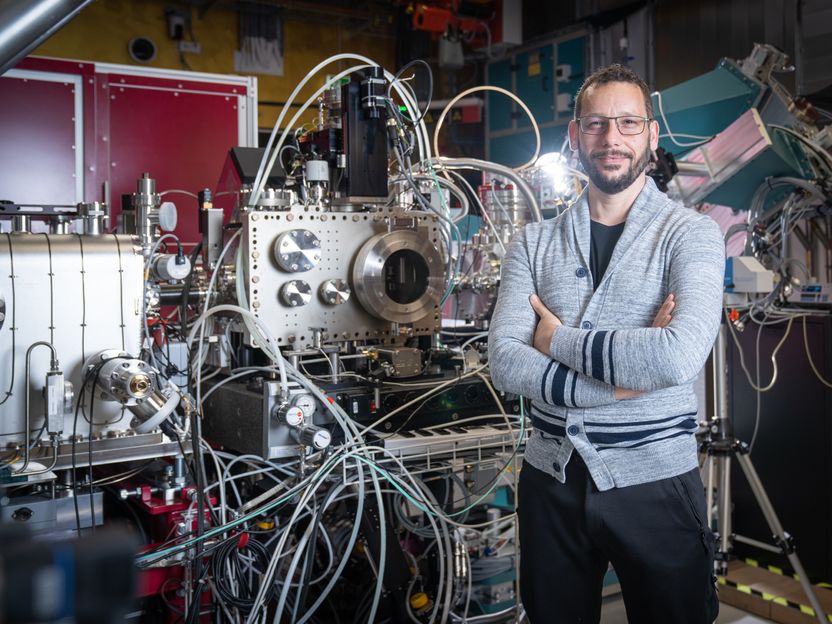
Cristian Svetina at the experiment station of the X-ray free-electron laser SwissFEL.
Mahir Dzambegovic, Paul Scherrer Institut
The structures on microchips are becoming ever tinier; hard disks write entire encyclopedias on magnetic disks the size of a fingernail. Many technologies are currently breaking through the boundaries of classical physics. But in the nanoworld, other laws apply – those of quantum physics. And there are still many unanswered questions: How does heat actually travel through a semiconductor material at the nanoscale? What exactly happens when individual bits are magnetised in a computer hard disk, and how fast can we write? There are still no answers to these and many more questions mainly because current experimental techniques cannot look deeply and precisely enough into the materials and because some processes take place far too quickly for conventional experimental methods. But if we want to push ahead with technical miniaturisation, we need to understand such phenomena at the atomic level.
The mix of methods makes the difference
Fresh impetus is now being brought to the matter thanks to a new method devised by PSI researcher Cristian Svetina, together with Jeremy Rouxel and Majed Chergui at EPFL in Lausanne, Keith Nelson at MIT in the USA, Claudio Masciovecchio at Fermi FEL in Italy, and other international partners. "The method is actually not new, though, and it has been used for decades in the optical regime with exceptional results," says Svetina, who is currently setting up the new Furka experiment station on the SwissFEL beamline Athos at PSI. What is special, he says, is the combination and extension of known methods from nonlinear laser physics, but using X-ray light from the new X-ray free-electron laser SwissFEL. This combination is both new and surprising. Several attempts have been made in the past by many groups around the world but without success. It has even been questioned whether such novel experiments could be successfully conducted at all at the high energies of X-rays. The team at PSI has proven: Yes, it can be done.
At its core, this is a method called transient grating spectroscopy. Spectroscopy is a proven set of methods used by physicists to obtain information about a material, such as the chemical elements and compounds it consists of, its magnetic properties, and how atoms move within it. In the particular variant called transient grating spectroscopy, the sample is bombarded with two laser beams that create an interference pattern. A third laser beam is diffracted at this pattern, creating a fourth beam that contains the information about the sample's properties.
Looking beneath the surface
The term laser is always used to describe light in the visible or infrared range of the wavelength spectrum. Therefore lasers can look inside a sample only with resolution limited to hundreds of nanometres. To go beyond this, X-rays are needed. Researchers at PSI have now succeeded for the first time in making transient grating spectroscopy accessible to an X-ray laser, using very hard X-rays with an energy of 7.1 kiloelectronvolts, which corresponds to a wavelength of 0.17 nanometres, or about the diameter of medium-sized atoms. The advantage: For the first time, it is possible to look inside materials with a resolution down to individual atoms as well as with ultrashort exposure times of fractions of femtoseconds (one millionth of a billionth of a second), which even allows videos of atomic processes to be recorded. In addition, the method is element-selective, meaning that one can selectively measure specific chemical elements in a mixture of substances. The method complements well established techniques such as inelastic neutron and X-ray scattering, adding better resolution in terms of both time and energy.
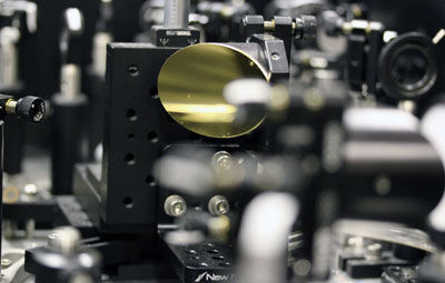
In practice, the experimental setup looks like this: SwissFEL sends a beam with a diameter of 0.2 millimetres, consisting of ultrashort X-ray pulses, onto a transmission phase grating made of diamond, which looks like a fine comb under the microscope. Diamond is used because it is not destroyed even by high-energy X-rays. It was made especially for this experiment by Christian David of the Laboratory for Micro and Nanotechnology at PSI. The spacing between the teeth of the comb is two micrometres, but this can go down to nanometres if needed. They break the X-ray beam into fine partial beams that overlap behind the grating, thus creating the transient grating diffraction pattern. Behind the grating, one-to-one images of the grating can be observed, repeated at regular intervals – so-called Talbot planes. If you place a sample in one of these planes, some atoms within it become excited, just as if it was sitting at the location of the grating. Only the atoms that "see" the X-rays in this periodic modulation are excited, while the neighbours that don’t experience the irradiation remain in the ground state. This is the chief attraction of the method, since it enables researchers to selectively excite characteristic domains of interest.
Camera with flash
Excitation of the atoms alone, however, does not provide any information. For this, a kind of camera with a flash is needed to briefly expose the sample. In transient grating spectroscopy, this is done by a laser that targets the sample at an angle and shoots images with a minimal time delay to the X-ray beam from SwissFEL. The information comes out of the back of the sample and hits a detector that records the image. Initial experiments have shown one advantage of the method: It does not produce any unwanted background signal. "If the atoms are excited, you see a signal; if they are not excited, you see nothing," Svetina explains. This is extremely valuable when measuring samples that emit only weak signals and that cannot be seen with other techniques where a background obscures the signal.
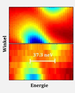
The fact that Cristian Svetina and his team have managed to do what other researchers have not is due to the creativity and patience of the protagonists. "We proceeded step by step and did not want to try everything at once," says the physicist. Five years ago the researchers started experimenting at FERMI FEL with optical light and extended it to extreme ultraviolet light before moving on to X-rays at PSI. Here, instead of examining "real" samples right away, they used gold foils to test whether the energy was sufficient to excite atoms. They succeeded in burning the lattice pattern from a Talbot plane into the foil. Svetina: "That's when we knew: If we can even print structures, we can excite atoms with lower intensity." With this the way was clear for the now successful experiment. Using a sample of bismuth germanate, the researchers were able to show that the method fulfilled all their hopes in terms of spatial and temporal resolution, measurement speed, and element selectivity.
Next goal: everything with X-rays
However, the researchers have not yet taken the final step. So far, only the beam that excites the sample is an X-ray beam. The flash of the camera still comes from a laser, so it is visible light. The pinnacle would be reached if that too were an X-ray beam. Svetina: "We want to take this final step in the course of the year." And they have additional support: SLAC's LCLS and the PULSE Institute, both at Stanford in California, the RIKEN SPring-8 centre in Japan, and DESY's FLASH in Germany have joined the collaboration team.
Original publication


Other news from the department science


Most read news


More news from our other portals


See the theme worlds for related content
Topic World Spectroscopy
Investigation with spectroscopy gives us unique insights into the composition and structure of materials. From UV-Vis spectroscopy to infrared and Raman spectroscopy to fluorescence and atomic absorption spectroscopy, spectroscopy offers us a wide range of analytical techniques to precisely characterize substances. Immerse yourself in the fascinating world of spectroscopy!