Visualization of the origin of magnetic forces by atomic resolution electron microscopy
Accelerating research and development on state-of-the-art materials such as magnets, semiconductors and quantum technology
Advertisement


The joint development team of Professor Shibata (the University of Tokyo), JEOL Ltd. and Monash University succeeded in directly observing an atomic magnetic field, the origin of magnets (magnetic force), for the first time in the world. The observation was conducted using the newly developed Magnetic-field-free Atomic-Resolution STEM (MARS). This team had already succeeded in observing the electric field inside atoms for the first time in 2012. However, since the magnetic fields in atoms are extremely weak compared with electric fields, the technology to observe the magnetic fields had been unexplored since the development of electron microscopes. This is an epoch-making achievement that will rewrite the history of microscope development.
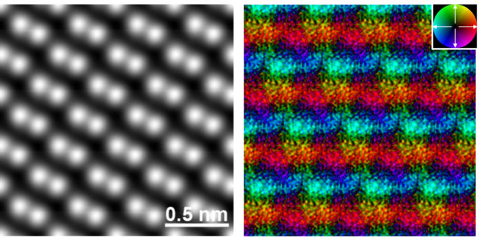
The atomic structure image (left) and corresponding magnetic field image (right). In the atomic structure image, Fe atoms are visualized as bright spots. In the magnetic field image, the color contrast indicates the magnetic field orientation and strength. The inset color wheel indicates how color and shade denote the magnetic field orientation and strength in the vector color map. The antiparallel magnetic fields on the adjacent Fe atomic layers are clearly observed, visualizing antiferromagnetic order in this crystal.
Naoya Shibata
Electron microscopes have the highest spatial resolution among all currently used microscopes. However, in order to achieve ultra-high resolution so that atoms can be observed directly, we have to observe the sample by placing it in an extremely strong lens magnetic field. Therefore, atomic observation of magnetic materials that are strongly affected by the lens magnetic field such as magnets and steels had been impossible for many years. For this difficult problem, the team succeeded in developing a lens that has a completely new structure in 2019. Using this new lens, the team realized atomic observation of magnetic materials, which is not affected by the lens magnetic field. The team’s next goal was to observe the magnetic fields of atoms, which are the origin of magnets (magnetic force), and they continued technological development to achieve the goal.
This time, the joint development team took on the challenge of observing the magnetic fields of iron (Fe) atoms in a hematite crystal (α-Fe2O3) by loading MARS with a newly developed high-sensitivity high-speed detector, and further using computer image processing technology. To observe the magnetic fields, they used the Differential Phase Contrast (DPC) method at atomic resolution, which is an ultrahigh-resolution local electromagnetic field measurement method using a scanning transmission electron microscope (STEM), developed by Professor Shibata et al. The results directly demonstrated that iron atoms themselves are small magnets (atomic magnet). The results also clarified the origin of magnetism (antiferromagnetism) exhibited by hematite at the atomic level.
From the present research results, the observation on atomic magnetic field was demonstrated, and a method for observation of atomic magnetic fields was established. This method is expected to become a new measuring method in the future that will lead the research and development of various magnetic materials and devices such as magnets, steels, magnetic devices, magnetic memory, magnetic semiconductors, spintronics and topological materials.
Original publication


Other news from the department science


