All-in-1 nanoparticle: A Swiss Army knife for nanomedicine
Advertisement


nanoparticles are being developed to perform a wide range of medical uses – imaging tumors, carrying drugs, delivering pulses of heat. Rather than settling for just one of these, researchers at the University of Washington have combined two nanoparticles in one tiny package. The result is the first structure that creates a multipurpose nanotechnology tool for medical imaging and therapy. The structure is described in Nature Nanotechnology.
"This is the first time that a semiconductor and metal nanoparticles have been combined in a way that preserves the function of each individual component," said lead author Xiaohu Gao, a UW assistant professor of bioengineering.
The current focus is on medical applications, but the researchers said multifunctional nanoparticles could also be used in energy research, for example in solar cells.
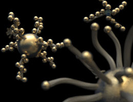
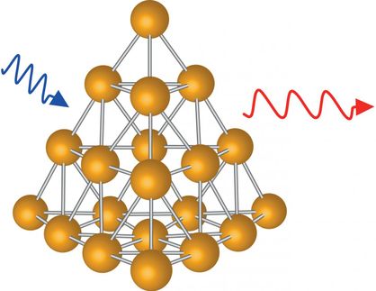
Quantum dots are fluorescent balls of semiconductor material just a few nanometers across, a small fraction of the wavelength of visible light (a nanometer is 1-millionth of a centimeter). Glowing gold nanoparticles are being developed for delivering drugs, for treating arthritis and for a type of medical imaging that uses infrared light. Gold also reradiates infrared heat and so could be used in medical therapies to cook nearby cells. But combine a quantum dot and a gold nanoparticle, and the effects disappear. The electrical fields of the particles interfere with one another and so neither behaves as it would on its own. The two have been successfully combined on a surface, but never in a single particle.
The paper describes a manufacturing technique that uses proteins to surround a quantum dot core with a thin gold shell held at 3 nanometers distance, so the two components' optical and electrical fields do not interfere with one another. The quantum dot likely would be used for fluorescent imaging. The gold sphere could be used for scattering-based imaging, which works better than fluorescence in some situations, as well as for delivering heat therapy.
The manufacturing technique developed by Gao and co-author Yongdong Jin, a UW postdoctoral researcher, is general and could apply to other nanoparticle combinations, they said.
"We picked a tough case," Gao said. "It is widely known that gold or any other metal will quench quantum dot fluorescence, eliminating the quantum dot's purpose."
Gao and Jin avoided this problem by building a thin gold sphere that surrounds but never touches the quantum dot. They carefully controlled the separation between the gold shell and the nanoparticle core by using chains of polymer, polyethylene glycol. The distance between the quantum dot core and charged gold ion is determined by the length of the polymer chain and can be increased with nanometer precision by adding links to the chain. On the outside layer they added short amino acids called polyhistidines, which bind to charged gold atoms.
The total diameter of the combined particle is roughly 15-20 nanometers, small enough to be able to slip into a cell. Incorporating gold provides a well-established binding site to attach biological molecules that target particular cells, such as tumor cells. Gold could also potentially amplify the quantum dot's fluorescence by five to 10 times, as it has in other cases. The gold sphere offers one further benefit. Gold is biocompatible, is medically approved and does not biodegrade. A gold shell could thus provide a durable non-toxic container for nanoparticles being used in the body, Gao said.


Other news from the department science


